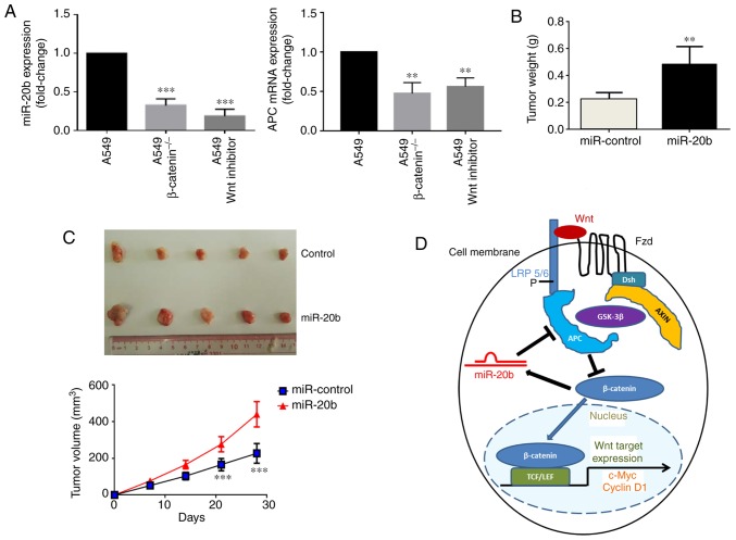
Figure 6.
miR-20b promotes tumor growth of H1975 in vivo. (A) Relative expression levels of miR-20b and APC in A549 (wild type), A549β-catenin(−/−) and A549 Wnt inhibitor cells determined by reverse transcription-quantitative PCR. **P<0.01 and ***P<0.001 vs. A549. Tumor (B) weight and (C) volume of xenograft tumors derived from nude mice (n=8 per group) injected with control or miR-20b-overexpressing H1975 cells after 28 days. As some of the samples were damaged during the excision process, 5 specimens from each group were presented. **P<0.01 and ***P<0.001 vs. miR-control. (D) Cell model of the miR-20b mechanism of action. miR, microRNA; APC, adenomatous polyposis coli; P, phosphate; Fzd, frizzled; Dsh, dishevelled; AXIN, axis inhibition protein; GSK-3β, glycogen synthase kinase 3β; TCF/LEF, T-cell factor/lymphoid enhancer-binding factor; LRP, lipoprotein receptor-related proteins.

