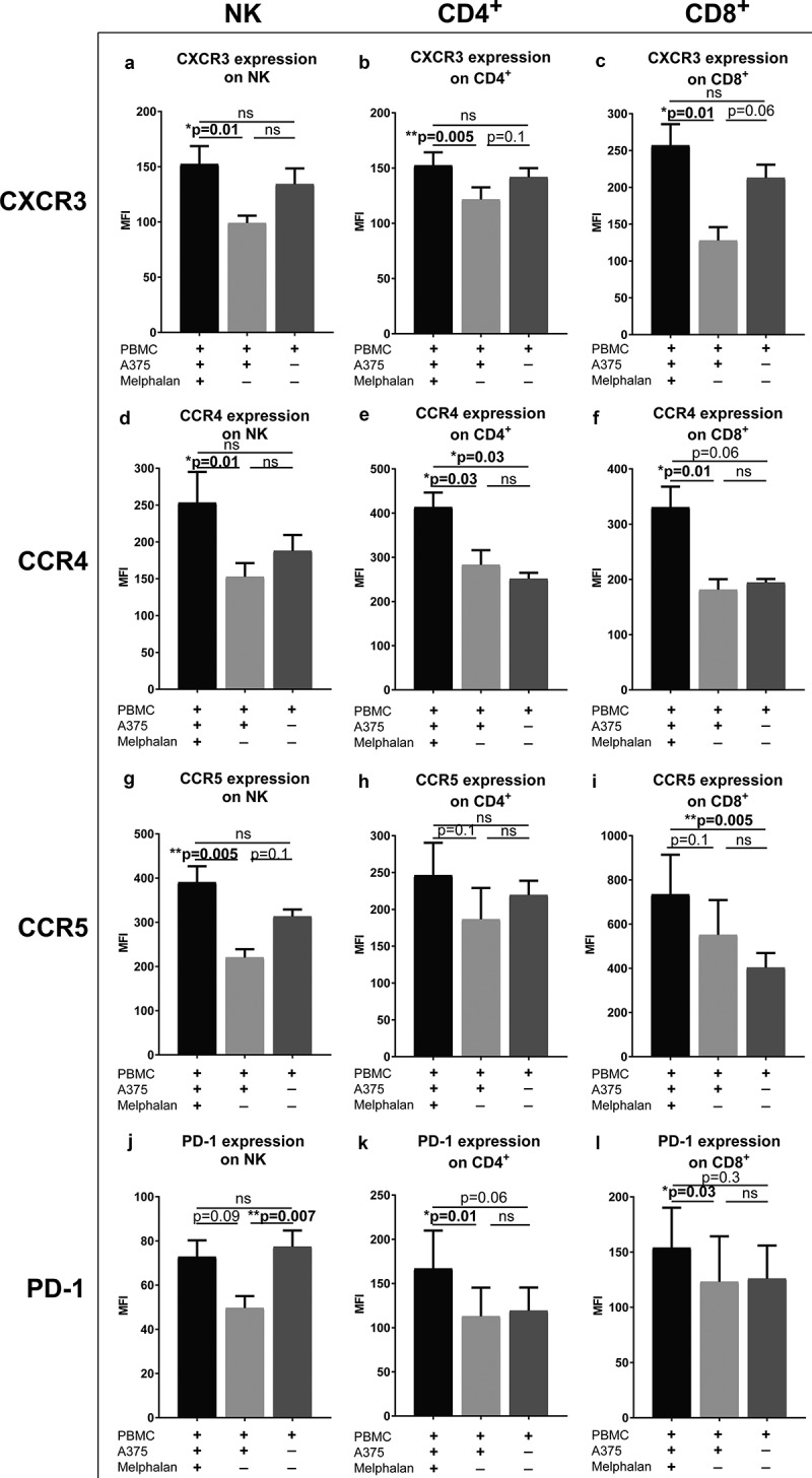
Figure 6.

Melphalan-exposed melanoma cells induce expression of receptors for ISG products on PBMCs.
PBMCs from healthy donors were cultured together with melphalan-exposed melanoma cells, non-exposed melanoma cells or were cultured alone. After 48 h, the PBMCs were transferred to new plates and were cultured in the absence of melanoma cells but presence of IL-2 for an additional 4 days. The expression of (a–c) CXCR3 (d-f) CCR4 (g–i) CCR5 and (j–l) PD-1 were measured on (a, d, g and j) NK cells (b, e, h and k) CD4+ T cells and (c, f, i and l) CD8+ T cells at the end of the culture (n = 6; paired Friedman test followed by Dunn’s multiple comparison test). MFI, Median Fluorescence Intensity. Data are presented as mean with SEM.
