
Figure 7.
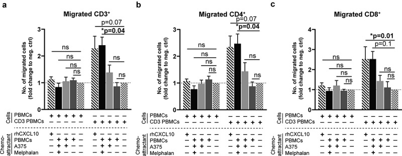
Activated T cells migrate toward supernatants from melphalan-exposed melanoma cells.
PBMCs from healthy donors cultured for 4 days in IL-2 containing medium in the presence or absence of an anti-CD3 antibody (CD3 PBMCs; clone: OKT3) were used for chemotaxis experiments. The number of (a) CD3+, (b) CD4+ and (c) CD8+ T cells that migrated toward supernatants from PBMC-melanoma co-cultures was determined after 4 h of migration. Recombinant human CXCL10 (rhCXCL10) was used as a positive control, while medium was the negative control. (n = 6; Friedman test followed by Dunn’s multiple comparison test). The fractions of migrated T cells are presented as fold change compared to the negative control. Data are presented as mean with SEM.