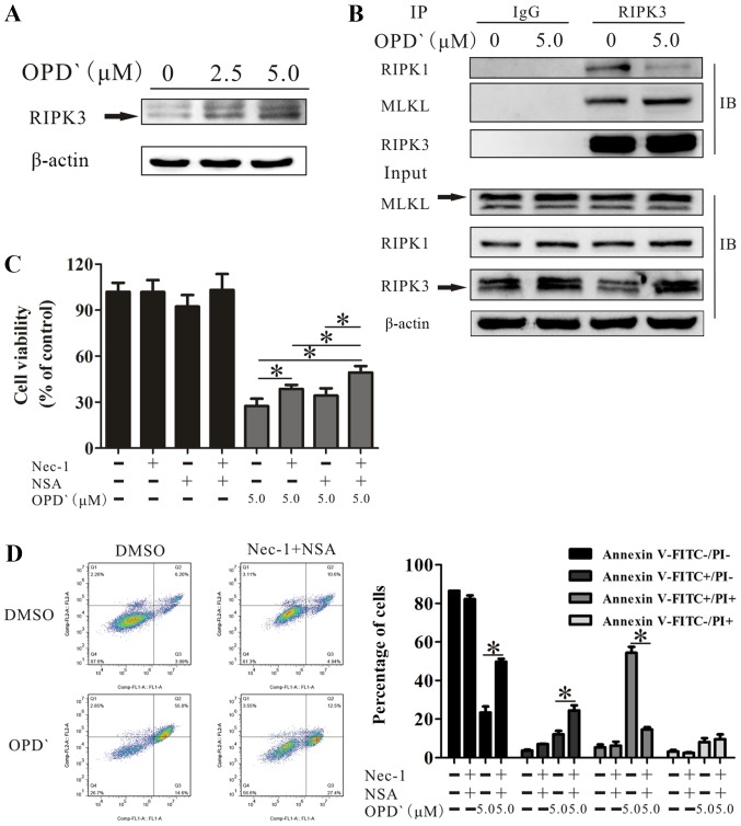
Figure 4.
OPD' induces necroptosis by multiple pathways involving RIPK1 and RIPK3/MLKL. (A) Western blotting was used to examine the protein expression of RIPK3 following treatment with OPD' for 6 h. (B) LNCaP cells were exposed to 5 µM OPD' for 6 h, then the protein lysates were immunoprecipitated using anti-RIPK3 antibodies. The target proteins RIPK1, MLKL and RIPK3 were examined by western blotting. (C) The Cell Counting Kit-8 assay was used to analyze the survival of LNCaP cells in response to OPD' following pre-treatment with Nec-1 and/or NSA (n=3). (D) Annexin V-FITC/PI staining was used to analyze the apoptotic rates of LNCaP cells exposed to OPD' for 24 h following combined pre-treatment with Nec-1 and NSA for 2 h. *P<0.05. OPD', Ophiopogonin D'; Nec-1, necrostatin-1; NSA, necrosulfonamide; RIPK, receptor-interacting serine/threonine-protein kinase; MLKL, mixed lineage kinase domain-like protein; PI, propidium iodide.

