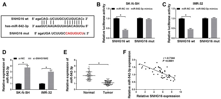
Figure 4.
SNHG16 acted as a sponge of miR-542-3p in NB cells. (A)The binding sites between SNHG16 and miR-542-3p were predicted by starBase database. The red sequence indicated that the mutation in TUG1 was at the predicted miR-542-3p binding sites. (B and C) The luciferase activity in SK-N-SH and IMR-32 cells co-transfected miR-542-3p mimics or miR-NC and SNHG16 wt or SNHG16 mut was determined with dual-luciferase reporter assay. (D) The expression of SNHG16 mRNA in SK-N-SH and IMR-32 cells transfected with si-SNHG16#2 or si-NC was detected using qRT-PCR. (E) QRT-PCR was used to assess the expression of miR-542-3p in NB tissues and corresponding adjacent normal tissues. (F) Pearson’s correlation analysis was performed for the assessment of the relationship between SNHG16 and miR-542-3p in NB tissues. *P<0.05.

