
FIGURE 1.
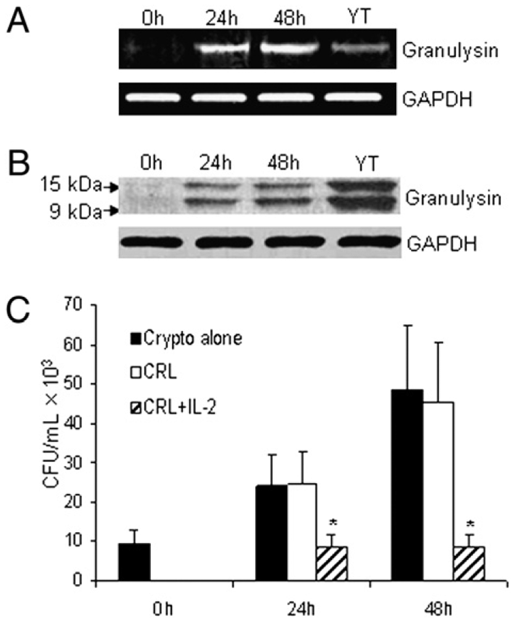
IL-2 promotes granulysin expression in CRL-2105 T cells. A, CRL-2105 T cells were stimulated for various times (0, 24, 48 h) with 100 U/ml IL-2 and compared with unstimulated YT cells. The expression of granulysin mRNA after IL-2 stimulation was assessed by RT-PCR, and GAPDH was used as an internal control. B, The time course of granulysin expression was also detected by Western blot using an Ab (519/GST) that detects both 15- and 9-kDa forms of granulysin. C, CRL-2105 T cells were treated with IL-2 and incubated with C. neoformans. The number of C. neoformans (CFU) was determined in each group as indicated. Results are expressed as mean ± SEM. Data are representative of three independent experiments. *p < 0.01 compared with the all other groups.