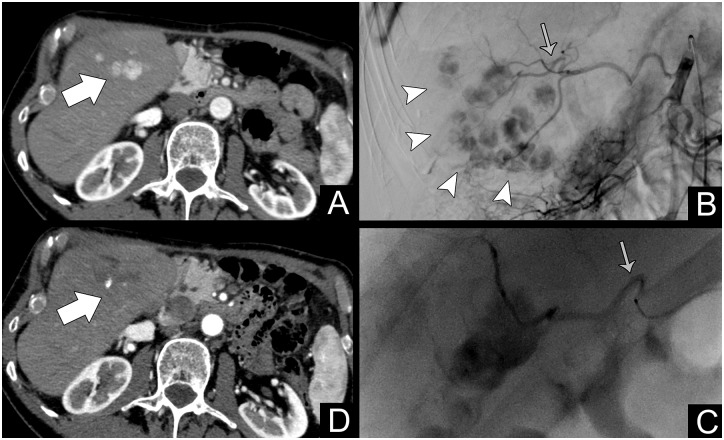
Fig 3. Patient #267, treated for multifocal HCC at 72 years (BCLC B).
A: Contrast-enhanced CT arterial phase showing a multifocal hypervascular HCC in the fifth segment (white arrow); B: Selective angiography of the right hepatic artery showing the extent of the disease (white arrowheads). The gray arrow points to the main feeding vessel; C: Superselective catheterization (gray arrow) and embolization of the main feeding vessel (with G-TAE); D: One-month follow-up showing complete devascularization with a small hyperdense spot representing residual Lipiodol (white arrow). The patient was still alive at the end of the study with a survival of 17.4 months.

