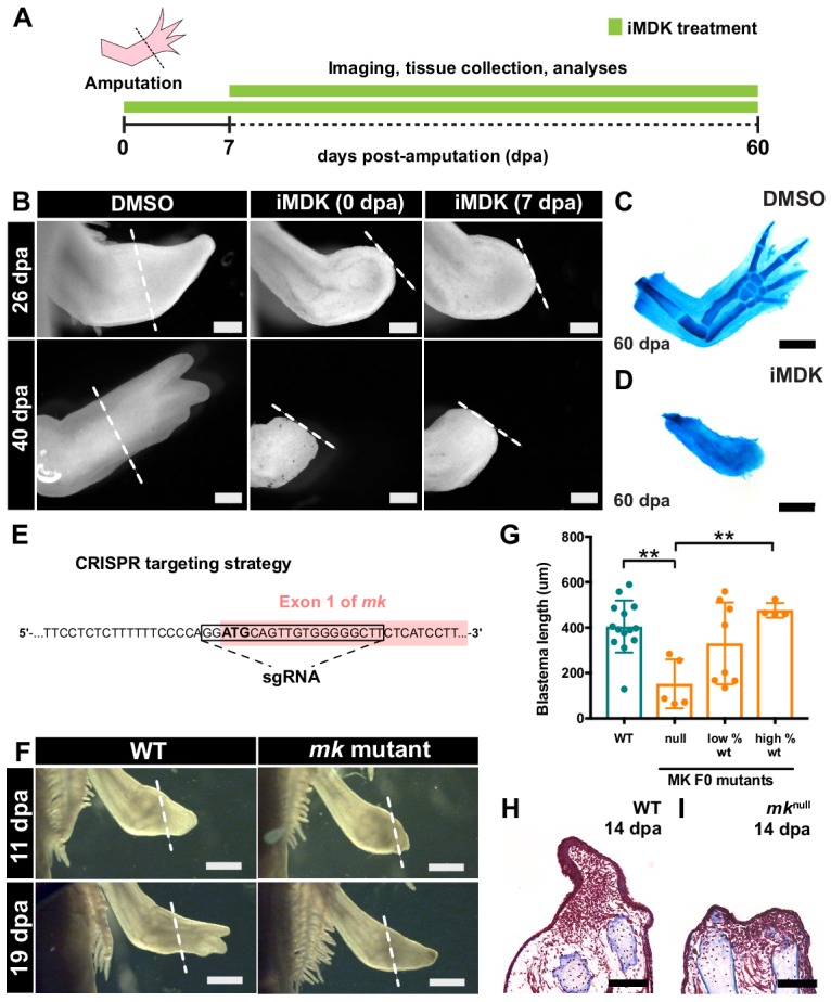
Figure 3. Chemical and genetic perturbations of mk impair limb regeneration.
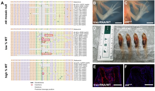
(A) Experimental design. (B) Brightfield images of DMSO- or iMDK-treated limbs (N = 4/4 iMDK-treated in each condition did not regenerate). (C–D) Alcian blue staining of DMSO- or iMDK-treated limbs at 60 dpa. (E) CRISPR strategy to target the start codon of mk to generate mutants. Control animals were generated using a non-targeted tracrRNA/cas9 complex and were unmodified at the mk locus. (F) Brightfield images of regenerating limbs from control animals or mk F0 mutants. (G) Quantification of blastema length at 14 dpa in control or mk mutant limbs. The severity of the delay in regeneration segregated based on genotype of the animal as either control (mkWT), mosaic null (mknull), or partially modified (low or high % WT alleles) (N = 14 control mkWT, 5 mknull, 8 low % WT, 4 high % WT). Graph is mean ± SD. (H–I) Representative images of picro-mallory stained sections of regenerating limbs in control animals or mk null mutants. Example genotyping analyses and mk null mutant immunofluorescence validation can be found in Figure 3—figure supplement 1. **p<0.005, two-tailed unpaired Student’s t-test was employed. Each N represents one limb from a different animal. White dotted lines mark amputation plane. Scale bars, B-D, H-I: 500 µm, F: 1 mm. WT, wildtype, dpa, days post-amputation.


