Figure 5. Mk acts as a critical survival factor during AEC development.
(A) Representative TUNEL-stained sections from limbs of DMSO and iMDK-treated limbs. (B) Representative TUNEL-stained sections from limbs of regenerating mkWT control or mknull mutants. (C) Quantification of the % TUNEL+ nuclei in the wound epidermis of DMSO or iMDK-treated limbs limbs at 5 dpa (N = 6 DMSO, 6 iMDK) or 11 dpa (N = 8 DMSO, 6 iMDK). (D) Quantification of the percentage of TUNEL+ nuclei in mkWT control and mknull mutants at 7 dpa (N = 12 mkWT, 8 mknull) and 10 dpa (N = 12 mkWT, 13 mknull). Asterisks mark auto-fluorescent bone. Each N represents a limb from a different animal. Quantification of levels of blastemal cell death in DMSO/iMDK-treated and mkWT/mknull regenerating limbs can be found in Figure 5—figure supplement 1. Two-tailed unpaired student’s t-tests were used for statistical analysis. Graphs are mean ± SD. *p<0.05, **p<0.005. Scale bars: 100 µm. dpa, days post-amputation.

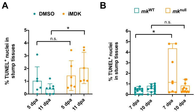
Figure 5—figure supplement 1. Mk is not required for blastemal cell survival.