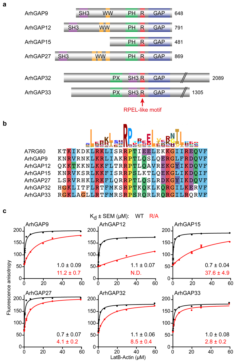
Figure 1. Two families of rhoGAPs contain an RPEL motif.
(a) Domain structure of ArhGAP12 and ArhGAP32 rhoGAP subfamilies. The RPEL-like motif is indicated in red. (b) Clustal X sequence alignment of the RPEL-like motifs of ArhGAP12/32 family GAPs with the RPEL motif of Nematostella vectensis A7RG60, aligned with the Pfam PF02755 HMM logo. (c) Fluorescence anisotropy analysis of LatB-actin binding to the FAM-conjugated RPEL peptides shown in (b), or derivatives in which the core RPEL arginine is replaced by alanine. Data were fitted by non-linear regression; data are means ± SEM, n=6 independent experiments. N.D., not determined. See Supplementary Figure 1 for related data. Source data for c are shown in Supplementary Table 1.

