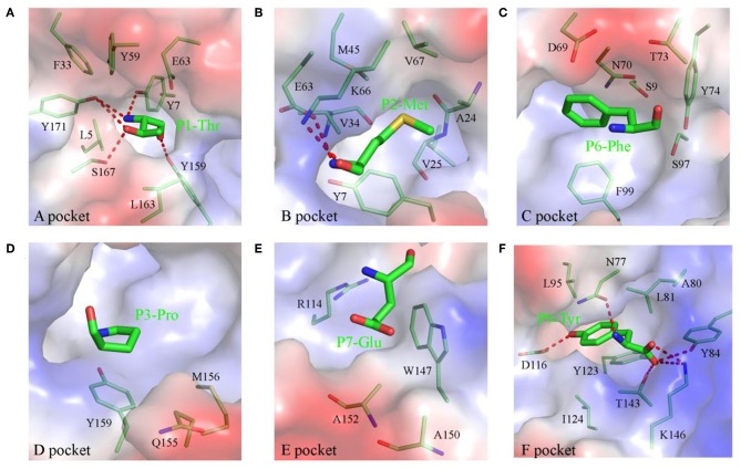
Figure 2.
Composition and polarities of the six pockets of pSLA-1*1502 bound to NSP9-TMP9 peptide. The pockets are shown in surface representation with their polarities colored as follows: red, negatively charged; white, non-polar; and blue, positively charged. The residues forming these pockets (light green) and the bound peptide (C, green; N, blue; O, red) are labeled. The hydrogen bonds between peptides and the red complex are shown with dashes. (A) Pocket A with residue P1 (Thr). (B) Pocket B with residue P2 (Met). (C) Pocket C with residue P6 (Phe). (D) Pocket D with residue P3 (Pro). (E) Pocket E with residue P7 (Glu). (F) Pocket F with residue P9 (Tyr).

