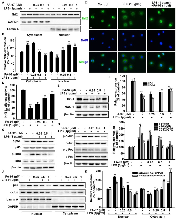
Figure 8.
Effect of FA-97 on Nrf2/HO-1 signaling pathway in vitro. RAW264.7 cells were pretreated with FA-97 (0, 0.25, 0.5, and 1 μM) for 24 h followed by LPS (1 μg/ml) stimulation for 2 h. (A) The expression of Nrf2 in cytosolic and nuclear extracts were determined by Western Blot. Lamin A and GAPDH were used as nuclear and cytoplasmic markers, respectively. (B) Densitometric analysis was performed to determine the relative ratios of Nrf2. GAPDH and Lamin A were used as nuclear and cytoplasmic markers, respectively. (C) RAW 264.7 cell slides were immune-stained with anti-Nrf2 (green) and DAPI (blue), and then the nuclear translocation of Nrf2 was observed by confocal laser-scanning microscope. (D) After transfected with ARE luciferase reporter plasmid, the Nrf2 transcription activity of RAW 264.7 cells was detected by luciferase activity assay. (E) The level of HO-1, NQO-1, and β-actin were detected by Western Blot. (F) Densitometric analysis was performed to determine the relative ratios of HO-1 and NQO-1. (G) Protein level of p-p65, p65, p-IκBα, IκBα, and β-actin in RAW 264.7 cells were detected by Western Blot. (H) The protein level of p-c-Jun, c-Jun, p-c-Fos, c-Fos, and β-actin in RAW 264.7 cells were detected by Western Blot. (I) Densitometric analysis was performed to determine the relative ratios of each protein. (J) The expression of p65 and c-Jun in cytosolic and nuclear extracts of RAW 264.7 cells were determined by Western Blot. Lamin A and GAPDH were used as nuclear and cytoplasmic markers, respectively. (K) Densitometric analysis was performed to determine the relative ratios of p65 and c-Jun. GAPDH and Lamin A were used as nuclear and cytoplasmic markers, respectively. Scale bars, 20 μm. The results are representative of three independent experiments. #P < 0.05, ##P < 0.01 compared with control group and *P < 0.05, **P < 0.01 compared with LPS-stimulated group.

