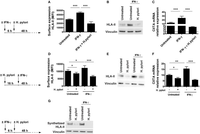
Figure 2.
Expression and transcriptional modulation of HLA-II in IFN-γ-activated macrophages infected by H. pylori. (A) Surface expression of HLA-II protein in human macrophages activated for 6 h with 20 ng/ml IFN-γ before the infection with H. pylori (MOI 10) for 48 h. Data are expressed as MFI ± SEM of three independent experiments, performed with cells from three different donors. (B) HLA-II total cell content in macrophages infected as in (A), evaluated by Western blot. Blot refers to a representative of three independent experiments, performed with cells from three different donors. (C) Relative expression of mRNA encoding CIITA in macrophages activated as in (A). Data were normalized to an endogenous reference gene (β-actin). Values at T0 cells were taken as reference and set as 1 (dotted line); the expression levels for treated cells were relative to the expression of T0 cells. Data are expressed as mean ± SEM of three independent experiments, performed with cells from three different donors. (D) Surface expression of HLA-II protein in human macrophages infected for 16 h with H. pylori (MOI 10) and treated with 20 ng/ml IFN-γ for 48 h. Data are expressed as MFI ± SEM of three independent experiments, performed with cells from three different donors. (E) HLA-II total cell content in macrophages treated as in (D), evaluated by Western blot. Blot refers to a representative of three independent experiments, performed with cells from three different donors. (F) Relative expression of mRNA encoding CIITA in macrophages activated as in (D). Data were normalized to an endogenous reference gene (β-actin). Values at T0 cells were taken as reference and set as 1 (dotted line); the expression levels for treated cells were relative to the expression of T0 cells. Data are expressed as mean ± SEM of three independent experiments, performed with cells from three different donors. (G) HLA-II synthetic rate in macrophages treated as in (A). At the end of stimulation, macrophages were labeled with the methionine analog AHA (L-Azidohomoalanine) for 2 h. Cells were lysed, and newly synthetized proteins were tagged with biotin alkyne. Proteins were pulled down with neutravidin agarose resin, processed for Western blot and developed for HLA-II. Blot refers to a representative of three independent experiments, performed with cells from three different donors. Significance was determined by Student's t-test. *p < 0.05; **p < 0.01; and ***p < 0.001.

