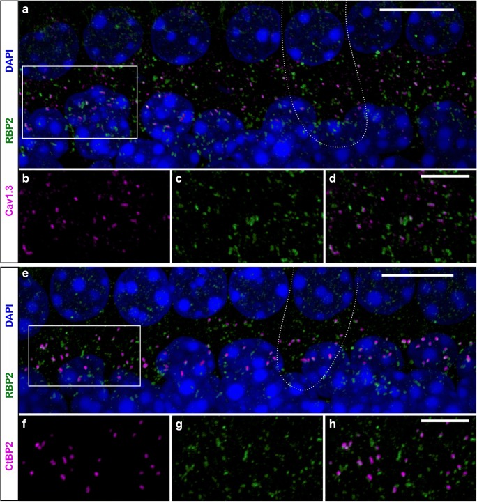
Fig. 3.
RBP2 co-localization with Cav1.3 at ribbon synapses in mouse IHCs. a–h Maximum intensity projection (MIP) of confocal stacks of whole-mount organs of Corti with stretches of 7–8 IHCs. a–d IHCs from the apical cochlear turn of a 4-week-old NMRI mouse co-immunolabeled for Cav1.3 and RBP2 demonstrate that almost every Cav1.3 cluster co-localized with RBP2 at the basolateral pole of the IHCs (a), which is shown in more detail in the enlargements of the box in a (b–d). e–h IHCs from the apical cochlear turn of a 4-week-old NMRI mouse co-immunolabeled for the ribbon synapse marker CtBP2 and RBP2 show that almost every ribbon co-localized with RBP2 at the basolateral pole (e), which is shown in more detail in the enlargements of the box in e (f–h). Nuclei stained in blue with DAPI are shown only in the merged images. The dotted lines in a and e outline the basolateral pole of one IHC in each specimen. Scale bars: a, e, 10 μm; d, h, 5 μm

