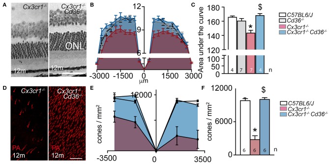
Figure 2.
CD36 deficiency prevents age-related rod and cone degeneration in Cx3cr1-knockout mice. (A) Micrographs, taken 1,000 μm from the optic nerve, of 12 month-old Cx3cr1−/− and Cx3cr1−/−Cd36−/− mice. (B) Photoreceptor nuclei rows at increasing distances (−3,000 μm: inferior pole, +3,000 μm: superior pole) from the optic nerve (0 μm) in 12 month-old mice. (C) Quantification of the area under the curve of photoreceptor nuclei row counts of 12 month-old mice of the indicated transgenic mouse strains (Mann Whitney wildtype vs. Cx3cr1−/− mice *p = 0.0024; Mann Whitney Cx3cr1−/− vs. Cx3cr1−/−Cd36−/− mice $p = 0.0024). (D) Micrographs, taken in the superior periphery of peanut agglutinin (staining cone segments, red) stained 12 month-old Cx3cr1−/− and Cx3cr1−/−Cd36−/− mice. (E) Cone density quantification on retinal flatmounts in peripheral and central, inferior and superior retina (−3,000 μm: inferior pole, +3,000 μm: superior pole, optic nerve: 0 μm) and their average (F) in 12 month-old mice of the indicated mouse strains (Mann Whitney wildtype vs. Cx3cr1−/− mice *p = 0.0022; Mann Whitney Cx3cr1−/− vs. Cx3cr1−/−Cd36−/− mice $p = 0.0022). ONL, outer nuclear layer; PNA, peanut agglutinin Scale bar (A,D) = 50 μm. n = number of replicates indicated in the graphs; replicates represent quantification of eyes from different mice of at least three different cages.

