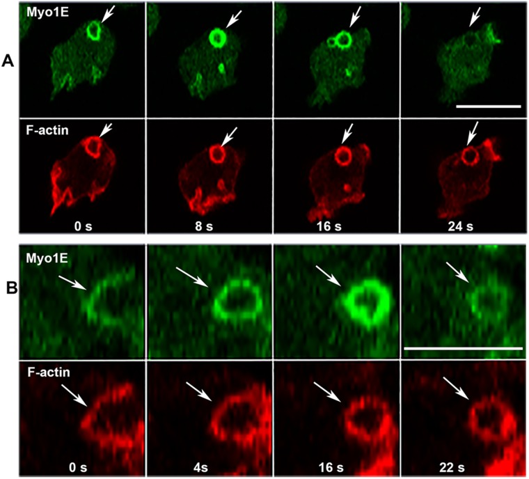
FIGURE 6:
Changes in Myo1E association with macropinocytic vesicles. (A) Myo1E dissociates from vesicle before F-actin. (B) Myo1E association with vesicle intensifies after vesicle internalization (16 s); images show high magnification of a single vesicle. Arrows point to the regions of interest in both A and B. Images are of live AX2 cells. Bars are 10 μm. Two independent Myo1E transfections were done and two independent experiments were performed for each transfection. The images are representative of more than 100 cells observed.

