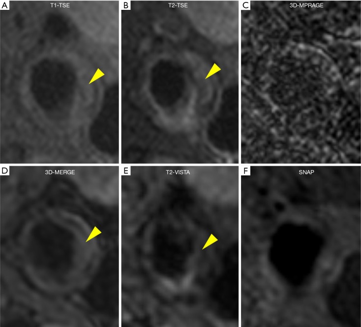
Figure 5.
Reference (A,B,C) and 3D (D,E,F) multi-contrast vessel wall images from a male patient with LRNC (yellow arrow). T1-TSE, T1-weighted turbo spin echo; T2-TSE, T2-weighted turbo spin echo; 3D-MPRAGE, three-dimensional magnetization-prepared rapid acquisition gradient-echo; 3D-MERGE, 3D motion-sensitized driven equilibrium prepared rapid gradient echo; T2-VISTA, T2-weighted volumetric isotropic turbo spin echo acquisition; SNAP, simultaneous non-contrast angiography and intraplaque hemorrhage.

