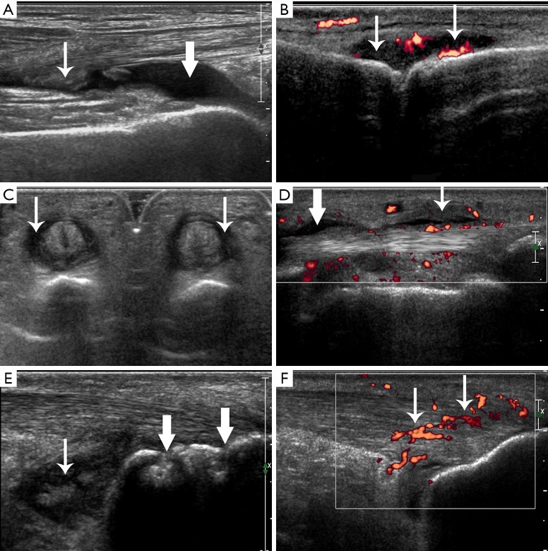
Figure 1.
Ultrasonic appearances of joint, tendon, enthesis, and bursa changes. (A) Joint effusion (crude arrow) and synovial thickening (fine arrow) in a left knee in a non-PsA patient. (B) Joint synovial thickening with PD signals of grade 2 (arrows) in a right metacarpophalangeal joint in a PsA patient. (C) Tendon sheath synovial thickening (arrows) in digital flexor tendons of a left hand in a PsA patient. (D) Tendon sheath effusion (crude arrow) and tendon sheath synovial thickening with PD signals (fine arrow) in a digital flexor tendon of a right hand in a PsA patient. (E) Enthesis bone erosion (crude arrows) in a left Achilles tendon and bursa synovial thickening (fine arrow) in the retrocalcaneal bursae in a PsA patient. (F) Enthesis thickening, hypoechogenicity with PD signals (arrows) in a right Achilles tendon in a PsA patient. PsA, psoriatic arthritis; PD, power Doppler.

