-
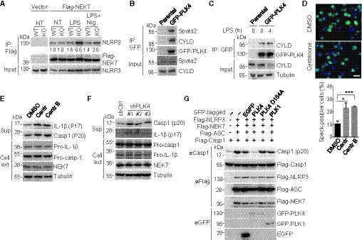
A
WT and Spata2 KO iBMDMs stably expressing an empty vector or a vector encoding Flag‐NEK7 were untreated (NT), primed with LPS for 4 h, or primed with LPS for 4 h followed by stimulation with Nigericin for 30 min. Flag‐NEK7 was immunoprecipitated, and the association of endogenous NLRP3 was assessed by immunoblotting. Relative levels of NLRP3 in immunoprecipitates were normalized to Flag‐NEK7 and shown below.
-
B
Co‐immunoprecipitation analysis of PLK4 interaction with endogenous Spata2 and CYLD in parental HEK293 cells and HEK293 cells stably expressing GFP‐PLK4.
-
C
iBMDMs stably expressing GFP‐PLK4 and parental iBMDMs were unstimulated or stimulated with 0.5 μg/ml LPS for 4 h. The PLK4 interaction with endogenous CYLD was analyzed by co‐immunoprecipitation with anti‐GFP antibody.
-
D
Confocal microscopy analysis of ASC specks in iBMDMs stably expressing GFP‐ASC, pretreated for 2 days with DMSO and two different PLK4 inhibitors, Centrinone (150 nM) and Centrinone‐B(500 nM), primed with LPS for 4 h, and stimulated with Nigericin for 30 min. GFP‐ASC specks were imaged and quantified (> 500 cells counted). Scale bar, 10 μm. Bars and error bars represent the mean ± SD of triplicate experiments. *P < 0.05; ***P < 0.001. Statistical analysis was performed using unpaired two‐tailed Student's t‐test.
-
E, F
Immunoblot analysis of the indicated proteins in cell supernatants (Sup) and cell extracts (Cell ext) of LPS‐primed and Nigericin‐stimulated BMDMs that were pretreated for 2 days with PLK4 inhibitors, Centrinone (150 nM) and Centrinone‐B (500 nM) (E), or of LPS‐primed and Nigericin‐stimulated iBMDMs stably expressing a control shRNA or three different PLK4‐specific shRNAs (F).
-
G
Immunoblot analysis of active caspase‐1 (p20) generation in HEK293 cells that were transfected with inflammasome components (Flag‐tagged NLRP3, NEK7, ASC, and caspase‐1) along with EGFP, GFP‐tagged PLK4, PLK4 kinase dead mutant (D154A), or PLK1 and stimulated with 10 μM Nigericin for 1 h 24 h post‐transfection.
Data information: For all Immunoblotting data, molecular weights in kDa are indicated to the left.