-
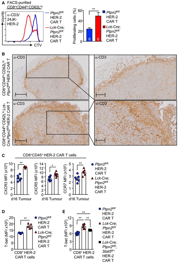
A
CD8+CD44hiCD62Lhi HER‐2 CAR T cells generated from Ptpn2
fl/fl versus Lck‐Cre;Ptpn2
fl/fl splenocytes were stimulated with plate‐bound α‐CD3 and subsequently labelled with CTV and incubated with 24JK‐HER‐2 cells, and proliferation was determined by flow cytometry.
-
B
HER‐2‐E0771 tumours isolated from HER‐2 TG mice on day 10 after adoptive Ptpn2
fl/fl versus Lck‐Cre;Ptpn2
fl/fl HER‐2 CAR T‐cell transfer were analysed for CD3+ T‐cell infiltrates by immunohistochemistry. Scale bars: 500 μm (full size) and 200 μm (zoom).
-
C
HER‐2‐E0771 cells (2 × 10⁵) were injected into the fourth inguinal mammary fat pads of female HER‐2 transgenic (TG) mice. Six days after tumour injection, HER‐2 TG mice received total body irradiation (4 Gy) followed by the adoptive transfer of 6 × 10⁶ FACS‐purified CD8+CD62LhiCD44hi central memory HER‐2 CAR T cells generated from Ptpn2
fl/fl versus Lck‐Cre;Ptpn2
fl/fl splenocytes. Mice were injected with IL‐2 (50,000 IU/day) on days 0–4 after adoptive CAR T‐cell transfer. Lymphocytes were isolated from the tumours at day 16 post‐adoptive transfer and CXCR3, CXCR5 and CCR7 MFIs on CD45+CD8+ T cells determined by flow cytometry.
-
D, E
Intracellular T‐bet MFIs in CD8+ HER‐2 CAR T cells were determined by flow cytometry.
Data information: Representative flow cytometry profiles and results (means ± SEM) are shown from two independent experiments. In (A, C, D, E), significance was determined using 2‐tailed Mann–Whitney
U‐test. *
P < 0.05, **
P < 0.01.