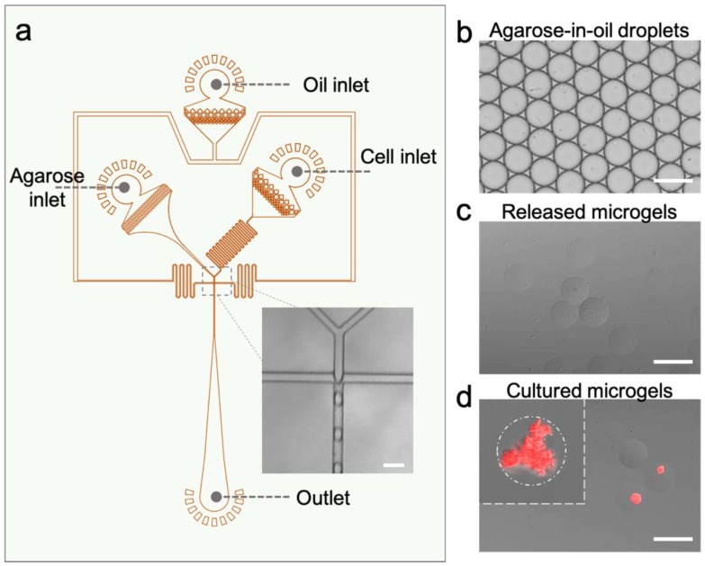
Figure 2.
Microfluidic manufacturing of the living lactam biosensors. (a) The layout of the droplet microfluidic device for the generation of agarose microgels with encapsulated engineered E. coli cells. (b,c) Microscopic images of (b) the as-generated agarose-in-oil droplets and (c) the released microgel beads suspended in an aqueous solution. (d) Stacked fluorescent micrograph showing microcolonies expressing mCherry proteins in the microgels induced by 50 mM caprolactam. Scale bars: 50 µm.

