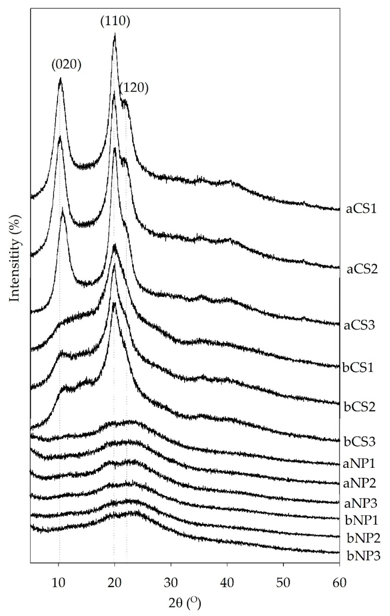
Figure 2.
X-ray diffraction patterns of α- and β-chitosan and α- and β-chitosan nanoparticles prepared from chitosan with different set of deacetylation degree (%DD) and molecular weight (MW) combinations, respectively. The analysis was conducted at 40 kV, 40 mA and 2θ with the scan angle from 5° to 60°. Where: a = α-chitosan, b = β-chitosan, CS = chitosan, NP = chitosan nanoparticles, 1 = low %DD and high MW, 2 = medium %DD and medium MW, and 3 = high %DD and low MW. (020), (110) and (120) represent the diffraction peak characteristic at 2θ ≈ 10°, ≈20°, and ≈21°, respectively.

