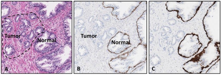
Figure 1.
Malignant primary prostate adenocarcinoma tissue sample. The first image shows the prostate tissue stained with H&E (A). The areas with tumor and normal prostate gland tissue are labeled. The H&E stained tissue slide was de-stained and anti-CK5/14 mouse monoclonal antibody cocktail was applied to determine feasibility of the proposed protocol (B). A sequential sample slide was stained with the same anti-CK5/14 marker using a standard protocol procedure (right panel), for comparison of stain intensity to initial de-stain/re-stain procedure results (C). [10× magnification].

