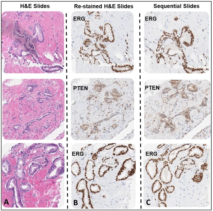
Figure 3.
Prostate cancer core needle biopsies (CNBs) sample H&E slides with PTEN and ERG IHC DAB stained slides. The first slide for each sample was H&E-stained (A). Each sample H&E was de-stained and re-stained with either PTEN or ERG antibody depending on the pathologist analysis for biomarker loss or positivity to demonstrate tumor heterogeneity (B). The additional sequential slides for each sample were stained with anti-PTEN antibody or anti-ERG antibody (C). Each sample H&E was de-stained and re-stained with either PTEN or ERG antibody depending on the pathologist analysis for biomarker loss or positivity to demonstrate tumor heterogeneity (10× magnification).

