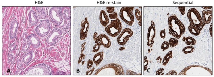
Figure 6.
The H&E stained slide archived 4+ years subjected to de-stain and re-stain procedure compared to the sequential sample slide. Initial H&E stained slide containing region of tumor (A). CK 8 &18 antibody re-stained slide retaining region of interest and exact architecture (B). Sequential slide comparison immunostained with CK 8 &18 exhibiting comparable stain intensity (C). [10× magnification].

