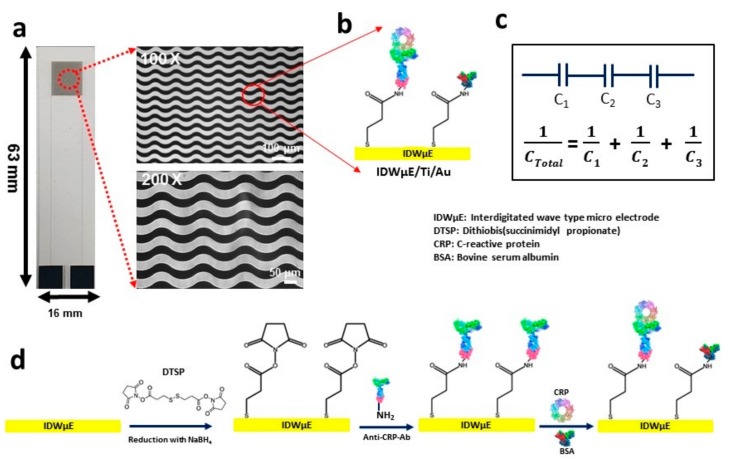
Figure 1.
Microscopic image of the IDWµE fabricated on a glass slide with dimensions of 16 mm × 63 mm × 1.1 mm. (a) The electrode array microscopic images at Low magnifications (100×) and high magnifications (200×) showing a 30 µm width for finger and spacing, respectively. (b) A schematic illustration of the DTSP functionalization and immobilization of anti-CRP-antibodies onto the IDWµE array. (c) The underlying working principle of the CRP immunosensor based on quantifying the total capacitance of the sensor after sequential formation of SAM, anti-CRP antibody, BSA, and CRP layers; (where C1 = CSAM, C2 = Canti-CRP-Ab, C3 = CCRP). (d) The sequential surface modification steps of the IDWµE array for immunosensing of CRP.

