Figure 10.
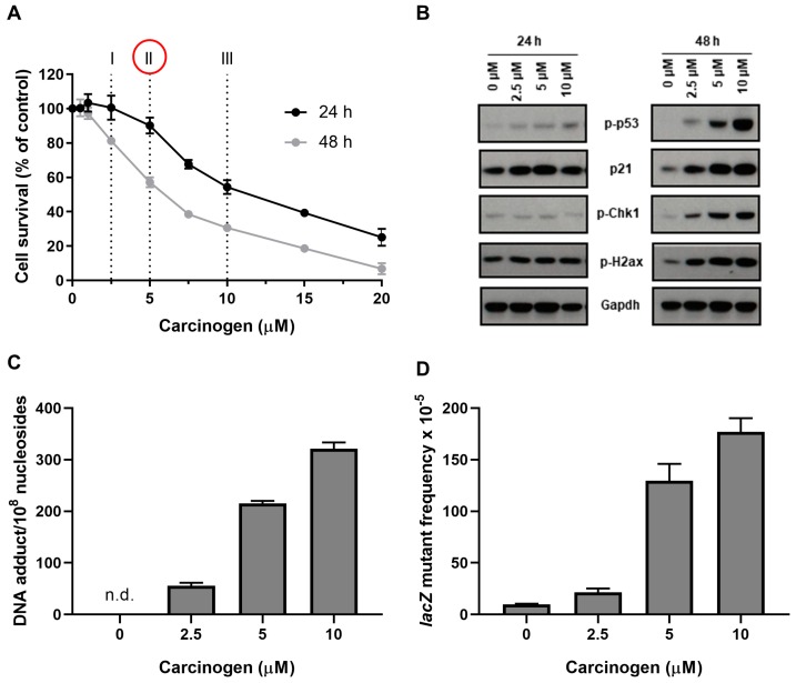
Optimisation of treatment conditions for the HIMA (simulated experiment). (A) Cell survival of primary HUFs after carcinogen treatment for 24 and 48 h. Cells treated with solvent served as control. Values represent mean ± SD (n = 3). Carcinogen concentrations I (~60–80% viable cells), II (~40–60% viable cells) and III (~20–40% viable cells) were used for testing DDR, DNA adduct formation and lacZ mutagenicity. (B) Expression of DDR proteins (i.e., p-p53, p21, pChk1, and pH2ax) by western blotting in primary HUFs after carcinogen treatment for 24 and 48 h. Cells treated with solvent served as control. Representative images of the western blotting are shown, and at least duplicate analysis was performed from independent experiments. Gapdh expression was used a loading control. (C) DNA adduct formation in primary HUFs after carcinogen treatment for 48 h. Cells treated with solvent served as control. Values represent mean ± SD (n = 4). n.d. = not detected. (D) Mutant frequency at the lacZ locus in primary HUFs after carcinogen treatment for 48 h. HUFs were treated with the indicated concentrations and then allowed to proliferate for 4 days to fix DNA damage into mutations. Cells treated with solvent served as control. LacZ mutant frequencies were calculated as the number of mutant colonies per number of recovered transformants. Values represent mean ± SD (n = 5). NOTE: Based on all assays conducted, we would recommend selecting carcinogen concentration II for the HIMA using a treatment period of 48 h (see red circle in panel A).

