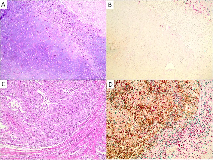
Fig. 3.
Representative images of different immune reaction and PD-L1 expressions in two invasive metaplastic breast carcinomas, as detected with anti-PD-L1 multiplex immunohistochemistry (anti-CD8 in green, anti-CD163 in red, and anti-PD-L1 in brown). a, b One invasive metaplastic carcinoma with no PD-L1 expression, only scattered CD163+ cells and very rare CD8+ cytotoxic T-cells in the peritumoral stroma. c, d One invasive metaplastic carcinoma with strong PD-L1 expression in tumor cells and stromal cells, diffuse CD163+ cells and CD8+ cytotoxic T-cells in tumoral stroma and peritumoral stroma. Magnification: × 100

