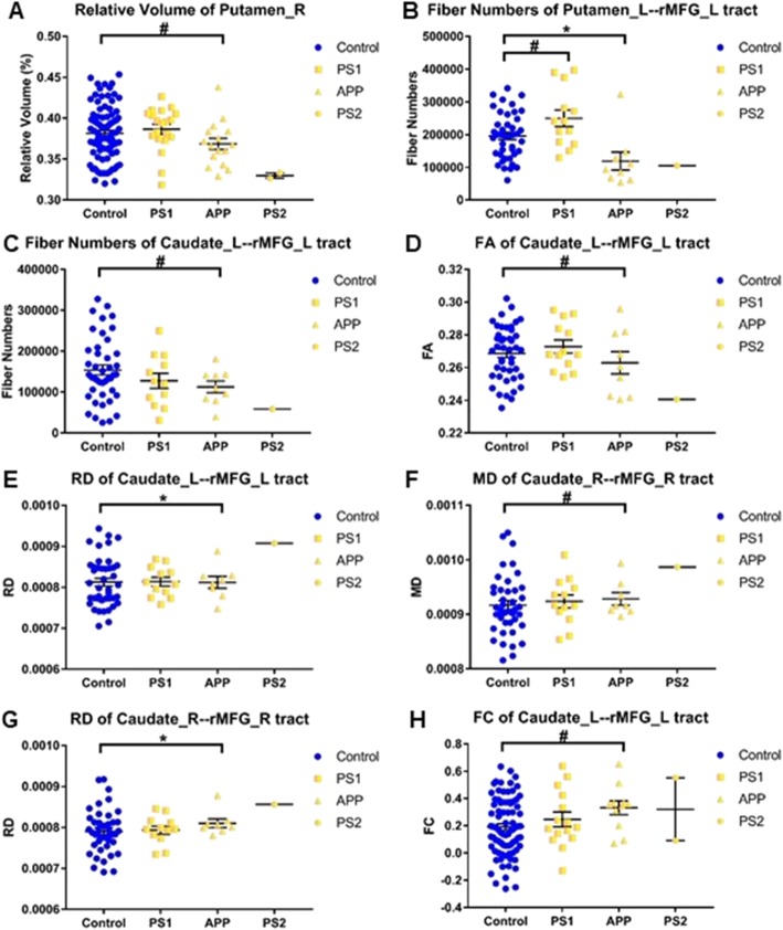
Fig. 5.
Effects of gene mutation on the imaging measures of neural circuits. a) relative volume of right putamen, b) fiber numbers of left putamen-rMFG tract, c) fiber numbers of left caudate-rMFG tract, d) FA of left caudate-rMFG tract, e) RD of left caudate-rMFG tract, f) MD of right caudate-rMFG tract, g) RD of right caudate-rMFG tract, h) FC of left caudate-rMFG tract. PS1 and APP mutation subjects without symptoms were compared with the control group, respectively. PS2 mutation subjects were listed for reference though not compared with the control group due to the small sample size. The bars indicate mean (SD). # 0.025 < P < 0.05, * 0.01 < P < 0.025

