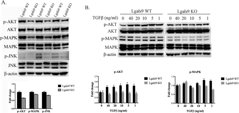
Fig. 4.
Effect of galectin-9 on the AKT, MAPK, and JNK pathways in vivo and in vitro. a Protein and phosphorylation levels in the lung tissues of galectin-9 wild-type (WT) and knockout (KO) mice treated with bleomycin for 4 weeks analyzed by western blotting for p-AKT, AKT, p-MAPK, MAPK, p-JNK, JNK, and β-actin. Protein and phosphorylation levels were normalized to the level of β-actin. The relative fold change was compared with the WT group. Data are shown as the mean ± SD, n = 3. b Western blotting for p-AKT, AKT, p-MAPK, MAPK, and β-actin protein expression in primary lung fibroblast cells treated with the indicated concentrations of TGF-β for 24 h. Protein expression levels were normalized to that of β-actin. The relative fold change in expression levels of the galectin-9 WT and KO groups was respectively compared with that of TGF-β untreated cells. Data are shown as the mean ± SD, n = 3

