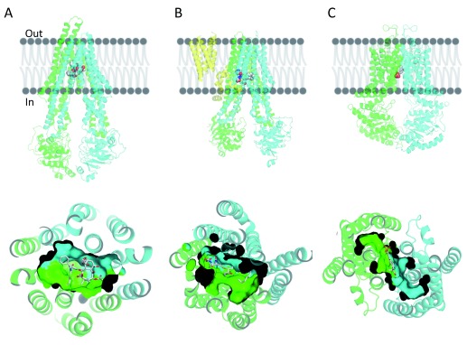
Figure 1.
The overall structure of ( A) taxol-bound P-glycoprotein (P-gp), ( B) leukotriene C4-bound multidrug resistance protein 1 (MRP1), and ( C) estrone-3-sulfate-bound ABCG2. Cartoon representation of the different transporters on the top panels and cartoon and surface representation of the binding cavity as observed from the cytosolic region in the bottom panels. TMD0 of MRP1 is colored in yellow, the N-terminal half of P-gp and MRP1 (transmembrane domain [TMD] 1 and nucleotide-binding domain [NBD] 1) are colored in green, and the corresponding C-terminal halves (TMD2 and NBD2) are colored in cyan. Each monomer of the homodimer of ABCG2 is colored in green or cyan. Ligands bound in the transmembrane region are shown in ball and stick format (gray, carbon; red, oxygen; blue, nitrogen).

