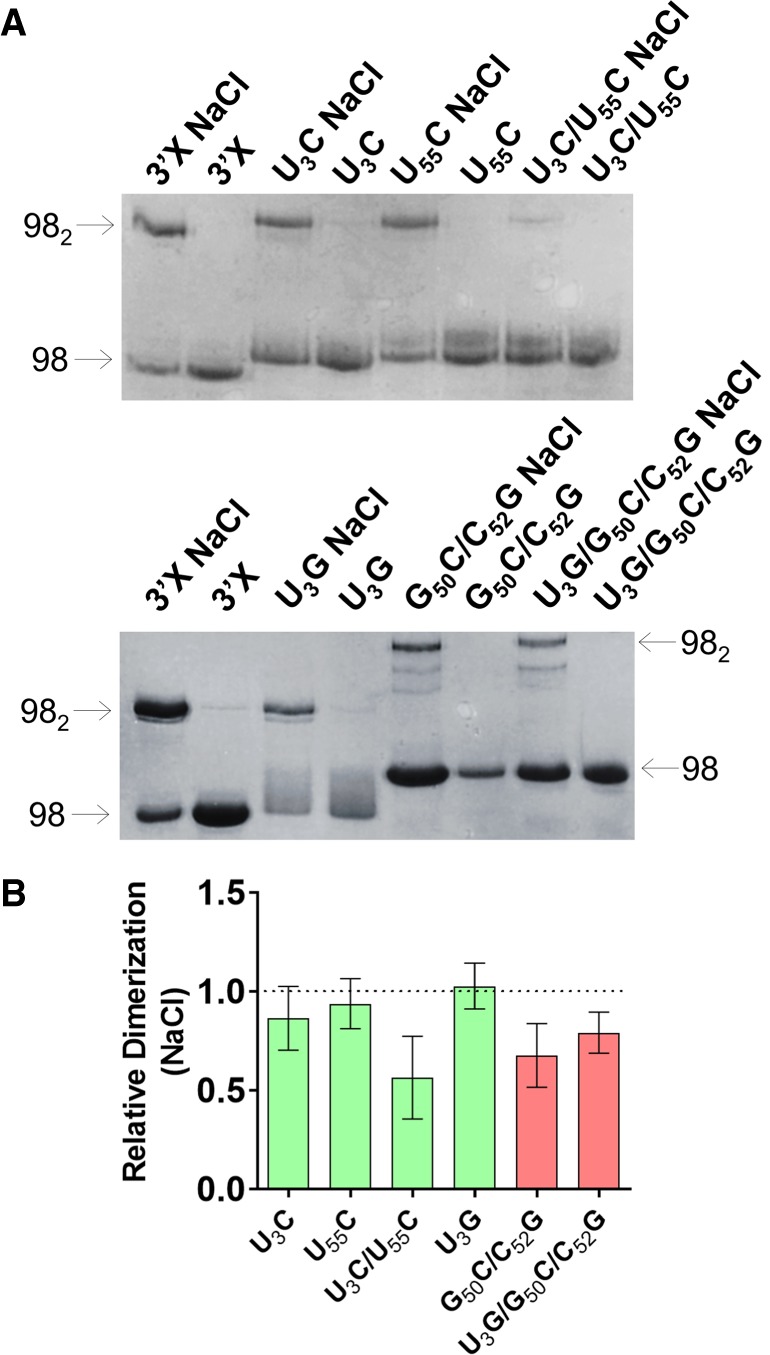
FIGURE 2.

Gel electrophoresis analysis of 3′X domain molecules as a function of ionic strength. (A) Representative native gels comparing the electrophoretic mobility of wild-type and U3C, U55C, U3C/U55C, U3G, G50C/C52G, and U3G/G50C/C52G mutant domain molecules, previously folded in the absence or presence of 100 mM NaCl. The arrows indicate the position of the monomer (98) and homodimer (982) species. (B) Quantification of the homodimerization yield at 100 mM NaCl of mutant molecules relative to the wild-type domain, which was assigned a value of 1. The bars represent the average and standard deviation of three independent experiments. Mutants experimentally verified to adopt the wild-type two-stem conformation are represented in green, whereas mutants adopting a different structure are indicated in red. Conditions: 10–40 µM RNA, TB running buffer.
