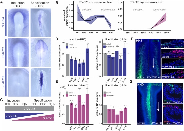
Figure 3.
TFAP2B and TFAP2C show complementary expression patterns and are required for induction and specification. (A) In situ hybridization for TFAP2A, TFAP2C, and TFAP2B in whole-mount chick embryos during induction (HH6) and specification (HH9). (B) Whole-embryo quantitative RT-PCR for TFAP2C and TFAP2B at six developmental stages (HH4–HH9) displays normalized mRNA expression relative to the housekeeping gene HPRT1. (C) TFAP2C and TFAP2B only partially recapitulate the expression pattern of TFAP2A. (D) Quantitative RT-PCR for induction genes MSX1, PAX7, ZIC1, and GBX2 (n = 6) and specification genes FOXD3, ETS1, SOX9, and SOX10 (n = 6), in control versus TFAP2C morpholino-treated sides of bilaterally electroporated embryos, represented as fold change compared with control. Phenotypes were surveyed at HH8− and HH9, respectively. (E) Quantitative RT-PCR for induction genes MSX1, PAX7, ZIC1, and GBX2 (n = 8) and specification genes FOXD3, ETS1, SOX9, and SOX10 (n = 8), in control versus TFAP2B DsiRNA-treated sides of bilaterally electroporated embryos, represented as fold change compared with control. Phenotypes were surveyed at HH8− and HH9, respectively. The neural marker SUFU was included as a control to ensure defects were neural crest specific. (F) Immunohistochemistry (whole-mount and transverse sections) for TFAP2C (green), as well as the induction marker PAX7 (red), in bilaterally transfected TFAP2C morpholino-treated embryos. Orientation of knockdown sections has been flipped for comparison. (G) Immunohistochemistry (whole-mount and transverse sections) for TFAP2B (green), as well as the specification marker SOX9, upon bilateral control versus TFAP2B DsiRNA treatment. (B,D,E) Error bars, SE. (HH) Hamburger Hamilton; (kd) knockdown; (n.s.) not significant. (*) P ≤ 0.05; (**) P ≤ 0.01; (***) P ≤ 0.001 (for number of embryos analyzed, see also Supplemental Table S1).

