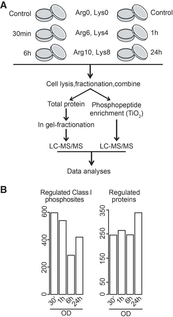
Figure 1.

Quantitative mass spectrometry revealed widespread changes during the initial period of osteoblast commitment. (A) Flow diagram of the quantitative proteomic and phosphoproteomic experiment. Human MSCs were labeled with the indicated combinations of arginine (Arg) and lysine (Lys) SILAC amino acids, induced to undergo osteoblast differentiation, and harvested at indicated time points. After cell lysis and fractionation, proteins were pooled, digested, and either analyzed by LC-MS/MS or enriched for phosphorylated species followed by LC-MS/MS analysis. (B) Numbers of significantly changed phosphorylation sites (left) and proteins (right) at indicated time points during osteoblastogenesis relative to undifferentiated cells.
