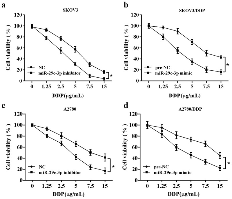
Figure 2.

Effect of miR-29c-3p on cell viability of DDP-treated ovarian cancer cells. (a) The DDP-sensitive cell line (SKOV3) was transfected with miR-29c-3p inhibitor followed by the DDP treatment with increasing concentrations (0, 1.25, 2.5, 5, 7.5, and 15 μg/mL) for 24 h. Cell viability was detected using the MTT assay. (b) The DDP-resistant cell line (SKOV3/DDP) was transfected with miR-29c-3p mimic followed by the DDP treatment with increasing concentrations (0, 1.25, 2.5, 5.0, 7.5, 15 μg/mL) for 24 h. Cell viability was detected using the MTT assay. (c) The DDP-sensitive cell line (A2780) was transfected with miR-29c-3p inhibitor followed by the DDP treatment with increasing concentrations (0, 1.25, 2.5, 5, 7.5, and 15 μg/mL) for 24 h. Cell viability was detected using the MTT assay. (b) The DDP-resistant cell line (A2780/DDP) was transfected with miR-29c-3p mimic followed by the DDP treatment with increasing concentrations (0, 1.25, 2.5, 5.0, 7.5, 15 μg/mL) for 24 h. Cell viability was detected using the MTT assay. Three independent experiments with biological repeats. *P < 0.05, vs. negative control (NC) or pre-NC.
