
Figure 1.
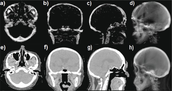
[(a)–(d)] Three orthogonal planes [(a)–(c)] of UTE‐MRI‐based bone image and an UTE‐MRI DRR (d) generated from a left lateral beam from a patient. [(e)–(g)] Three planes of CT image [(e)–(g)] and a CT‐based DRR (h) generated from a left lateral beam from the same patient.