
Figure 4.
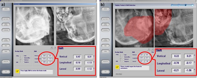
Patient setup example based on oblique image pairs. (a) 2D patient clinical setup result based on patient's simulation CT; (b) retrospective patient setup result by using patient's clinical ExacTrac images and the UTE‐MRI bone image. The calculated shifts in vertical, longitudinal, and laterals directions matched well between UTE‐MRI and simulation CT.