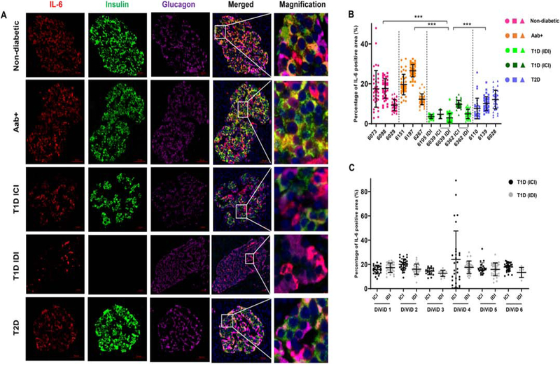
Figure 1. IL-6 is expressed on pancreatic beta cells and alpha cells and expression is reduced in insulin-deficient islets of donors with T1D.
(A) FFPE sections of human pancreata were stained with anti-IL-6 (red), anti-insulin (green), anti-glucagon (magenta) and Hoechst (blue). Representative images of islets from a non-diabetic donor (nPOD 6098), GAD auto-antibody positive (Aab+) donor (nPOD 6151), insulin-containing islet (ICI) of a T1D donor (nPOD 6362), insulin-deficient islet (IDI) of a T1D donor (nPOD 6195) and a donor with T2D (nPOD 6110) are shown. Images were acquired using Axioscan with a 20x objective. Scale bar – 50 μm. Systematic quantification of IL-6 staining was performed using Image Pro Premier software. (B) Percentages of the total islet area that stained positive for IL-6 in 3 non-diabetic (pink) donors, 3 auto-antibody positive (Aab+, orange) donors, insulin-containing islets (ICI, dark green) and insulin-deficient islets (IDI, light green) of 3 donors with T1D and 3 donors with T2D (blue). (C) Percentages of the total islet area positive for IL-6 staining in ICI (black) and IDI (gray) of recent onset T1D cases from the DiViD study. Graphs show mean ± SD of more than 30 islets per case. Each data point represents an islet. ***P<0.0001.

