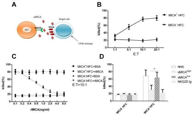
Figure 4.
sMICA inhibits the cytotoxicity of NK cells toward MICA+ target cells. (A) Schematic of components involved in the MICA-NKG2D pathway. NK effector cells were incubated with MICA+ target cells stained with CSFE dye. sMICA interferes with cell killing mediated by NK cells. (B) Target cells (5000 cells per assay) with (MICA+ HFC) or without MICA expressed human fibroblasts (MICA- HFC) were co-cultured with NKL at different E: T ratios. The mean percentages of dead cells from three replicate experiments are plotted. (C) The percent dead target cells in the presence of soluble rMICA, BSA at finally concentrations diluted from 0.1 to 8.0 ng/ml at E: T of 10:1. Plotted are means of triplicate experiments. (D) The percent dead target cells in the presence of serum from sMICAhigh patients (N=10) and from sMICAlow patients (N=10) at E:T of 10:1. Normal health serum (NHS) was used as negative control and soluble NKG2D-Ig at concentration at 1.0 μg/ml were used as maximum blocking controls. Plotted are means of three experiments for each tested (C and D).

