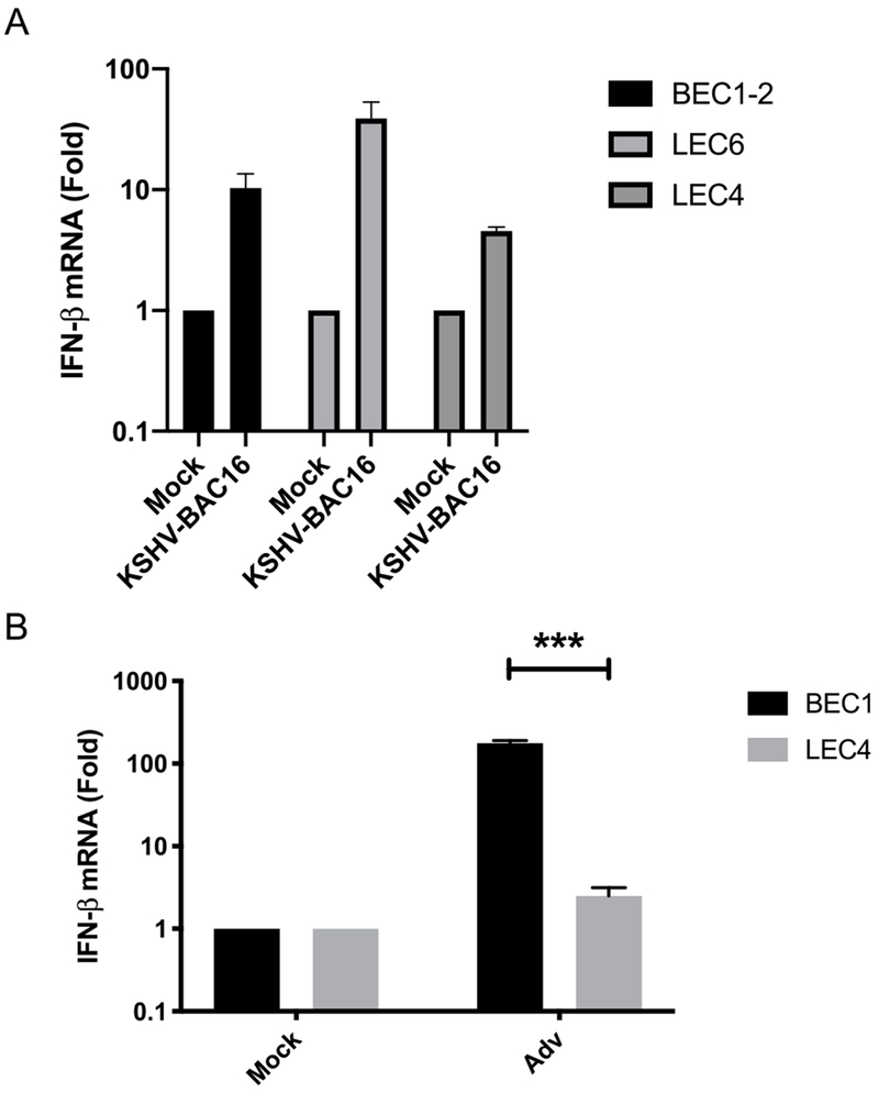
Fig. 1. KSHV induces minimal IFN-β expression in primary endothelial cells but LEC4 are defective for innate immune activation during Adv infection.
A) IFN-β mRNA was measured by RT-qPCR from BEC1-2, LEC4 and LEC6 that were infected with KSHV-BAC16 for 48 h. The relative amount of mRNA was normalized to tubulin mRNA in each sample, and fold change relative to mock was calculated (ΔΔct). B) IFN-β was measured by RT-qPCR from BEC1 and LEC4 that were infected with Adv for 48 h. The relative amount of mRNA was normalized as in (A). Data are shown as mean ± SEM from at least 3 biological replicates. ***P < 0.001; (Student’s t-test).

