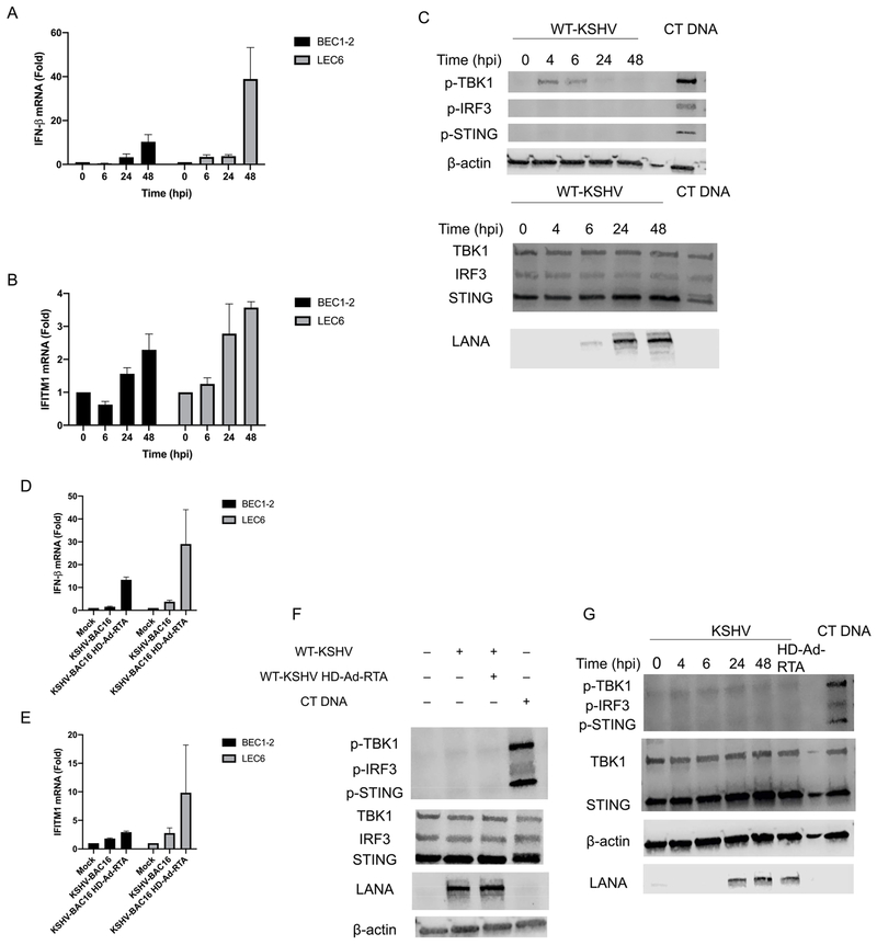
Fig. 5. KSHV induces minimal innate immune signaling and does not strongly activate STING-TBK1-IRF3 in primary endothelial cells.
(A) IFN-β or (B) IFITM1 mRNA was measured by RT-qPCR from BEC1-2 and LEC6 that were infected with KSHV-BAC16 and harvested at the indicated timepoints. The relative amount of mRNA for each gene was normalized as in Fig. 1. (C) BEC1 were either infected with KSHV or transfected with 1 μg/mL CT DNA and whole cell lysates were harvested at the indicated timepoints (3 h post transfection for cells transfected with CT DNA) and immunoblotted with the indicated antibodies. The top and bottom panels are the same lysate, run on different gels. (D) IFN-β or (E) IFITM1 mRNA was measured by RT-qPCR from BEC1-2 and LEC6 that were infected with either KSHV-BAC16 or KSHV-BAC16 + HD-Ad-RTA and harvested at 24 hpi. The relative amount of mRNA for each gene was normalized as in Fig. 1. (F) BEC1 cells were infected with WT-KSHV alone or WT-KSHV + HD-Ad-RTA and harvested at 24 hpi or transfected with 1 μg/mL CT DNA (3-h transfection). Whole cell lysates were immunoblotted with the indicated antibodies. (G) LEC6 were infected with WT-KSHV, WT-KSHV + HD-Ad-RTA or transfected with CT DNA and whole cell lysates were harvested at the indicated time points (24 h for WT-KSHV + HD-Ad-RTA cells and 3 h for CT DNA transfection) and immunoblotted for the indicated antibodies. Data are shown as mean ± SEM from at least 3 biological replicates.

