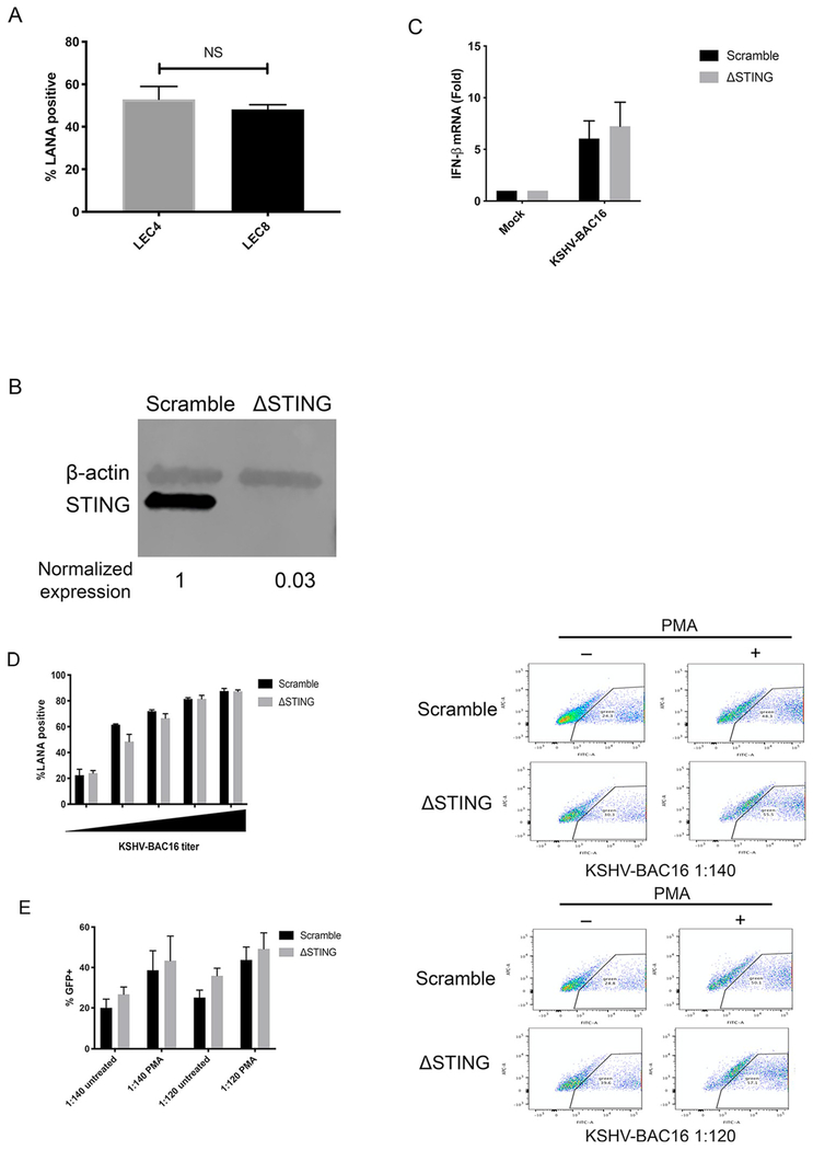
Fig. 6. STING does not reduce susceptibility during de novo infection or restrict spread of KSHV during lytic replication.
(A) LEC4 and LEC8 cells were infected with KSHV-BAC16 and infection rates were measured by immunofluorescence microscopy and quantifying the percent of LANA + cells in the culture at 48 hpi. (B) BEC1-3 were transduced with a lentivirus expressing Cas9 and a guide RNA targeting STING (ΔSTING) or a nontargeting (scramble) control and whole cell lysates were immunoblotted with the indicated antibodies. (C) Scramble and ΔSTING BEC3 from (B) were infected with KSHV-BAC16 and IFN-β mRNA was measured by RT-qPCR at 48 hpi as described earlier. (D) Scramble and ΔSTING BEC1-3 were infected with KSHV-BAC16 at different dilutions of virus and infection rates were measured by immunofluorescence microscopy and quantifying the percent of LANA + cells in the culture at 48 hpi. (E) Scramble and ΔSTING BEC3 were infected with KSHV-BAC16 at 2 different dilutions (1:140 and 1:120) for 4 h. Immediately following infection, cells were treated with PMA for 5 days. Cells were sorted by GFP by flow cytometry. Data are shown as mean ± SEM from at least 2 biological replicates.

