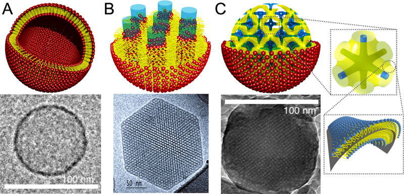
Figure 7.

Structure of three different lipid particles and their respective Cryo-EM images. A) Schematic of a liposome and the correspondent Cryo-EM image indicating a size of ca 100 nm in diameter. B) Hexosome representattion and the correspondent Cryo-EM image of a ca 200 nm particle. The Cryo-EM data is reprinted with permission from [86]. Copyright 2005 American Chemical Society. C) Cubosome schematics having a primitive bicontinuous cubic structure enclosed by a single lipid leaflet, where yellow represents the midplane of the lipid bilayer and blue the water channels. A single unit cell is also represented to the right of the cubosome schematic. The Cryo-EM shows a ca 100 nm cubosome. Reprinted with permission from [19]. Copyright 2018 American Chemical Society.
