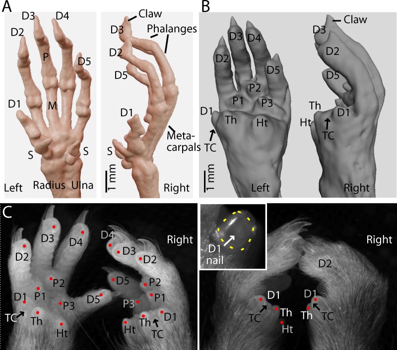
Fig 1. Anatomy of mouse D1.
(A) Micro-CT imaging of a pair of mouse hands showing the skeletal anatomy, with labeling of digits (D1-5) and sesamoid (S), metacarpal (M), and phalangeal (P) bones. (B) Soft-tissue rendering, showing the volar aspect, with labeling of the thenar (Th), hypothenar (Ht), and interdigital pads (P1-3), and the thumb cleft (TC) between the D1 and thenar pads. (C) Left: Macroscopic images of a pair of amputated mouse hands, showing major features of the volar aspect. Right: Same, but as a dorsal view with the hands positioned more naturally. Inset: Close-up view of the flat thumb nail on the left D1.

