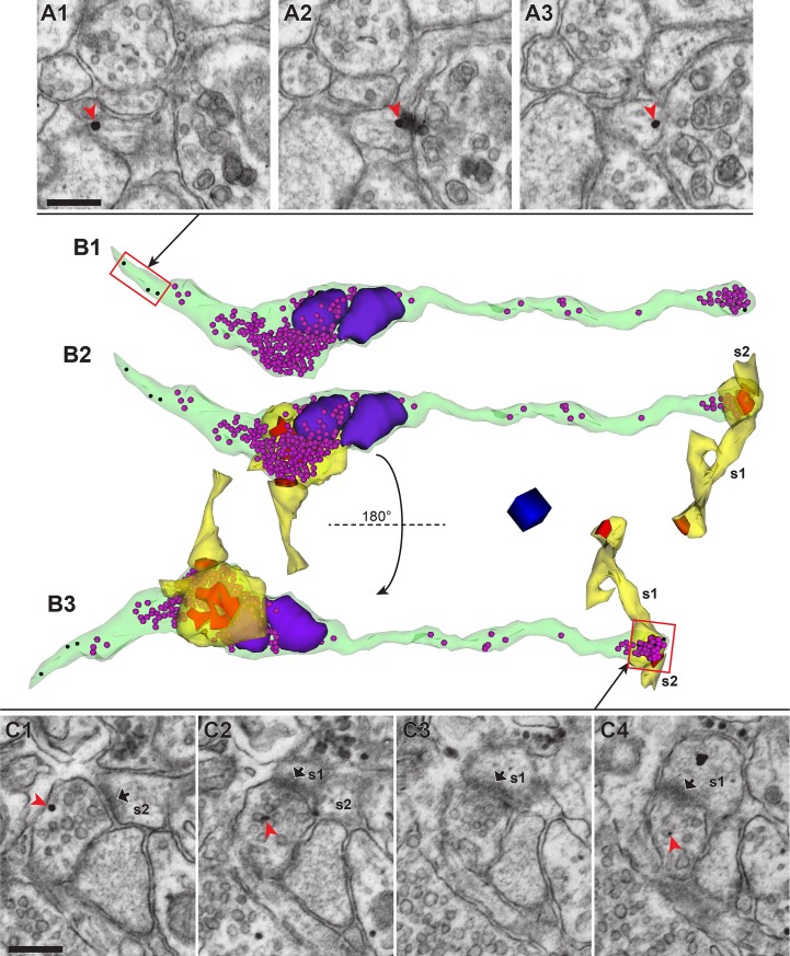
Fig 4. Post-embedding immunogold labeling for Alexa Fluor dyes deposited by mAPEX2-driven TSA reaction allowed for 3DEM identification of rAAV-targeted axons, while maintaining excellent ultrastructure.
(A1-A3) Three adjacent serial tSEM images of an axon containing immunogold labeling (red arrowhead). Scale bar = 250 nm. (B1) 3D reconstruction of the labeled axon (green) shown in A, which contained immunogold labels (black spheres) and synaptic vesicles (magenta spheres). Two mitochondria (purple) were associated with one of the two boutons. Red rectangle represents a portion of this axon shown in A1-A3. (B2) Same axon as B1, with reconstructions of spines (yellow) forming synapses (red) with this axon. Note, s1 was a branched spine with one of the heads forming a synapse with another axon. The second spine (s2) and its PSD could be reconstructed only partially because they were located at the end of the tSEM image series. Both axonal boutons are multi-synaptic, with each bouton forming synapses with two spines originating from different dendrites. (B3) Same as B2, rotated along the horizontal axis 180° to provide a different view of synapses and spines. One of the synapses at mitochondria-containing bouton was perforated (also see S1 Video). Red rectangle represents a portion of this axonal bouton shown in C1-C4. Scale cube = 250 nm per side. (C1-C4) Four adjacent serial tSEM images of the gold-labeled (red arrowhead) axonal bouton, forming synapses with two dendritic spines (s1 and s2; PSDs indicated by black arrows). Scale bar = 250 nm.

