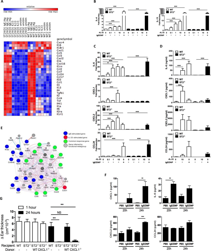
Fig 4. ST2-driven stimulation of basophils promotes a unique inflammatory profile in which basophil-derived CXCL1 is functionally important in their involvement in ADTI.
(A–C) BMBs from C57BL/6J mice were activated with IgE/DNP or different doses of mIL-33. Gene expression 4 hours after activation was analyzed by microarray (A) and real time RT-PCR (B, C). Protein production 24 hours after activation was determined by ELISA (D). (E) Network diagram generated from the microarray comparison of activated BMBs in (A) showing select genes altered by IgE-stimulation, IL-33-stimulation and shared by both stimuli. (F) Ear tissues from C57BL/6J mice undergoing the PCA model were collected at 20 and 24 hours after challenge and homogenized. IL-6, CXCL1, CXCL2 and CCL24 expression was determined by ELISA and normalized to total protein concentration. Data are from 3 individual mice at each time point (mean ± SEM). (G) WT or St2–/–underwent the PCA model. Some mice underwent repletion with WT or Cxcl1–/–BMBs 1 hour before challenge. Ear thickness was measured at 0, 1 and 24 hours after challenge. *P ≤ 0.05, **P ≤ 0.01, and ***P ≤ 0.001 (one-way ANOVA). Data are from at least 2 independent experiments, and mean ± SEM for n = 3–11 per group (B, C) n = 4–9 per group (D), n = 3 per group (F), and n = 6–10 per group (G) are displayed.

