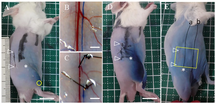
Fig 1. Surgical procedure for lymphatic ligation and the sectioning area for histological observation.
(A) Evans blue dye was injected into the hindlimb (yellow circle). A 1 cm incision was made 5 mm from the dorsal site in relation to the collecting LVs passing through the inguinal lymph node (LN) on the abdomen at 5 mm from the inguinal LN; scale bar of 1 cm. (B) The incised skin was peeled from the epimysium and the collecting LV (stained in blue color) passing through the inguinal LN was exposed; scale bar of 2 mm. (C) It was carefully tied at 2 points with 6–0 nylon sutures under a stereoscopic microscope; scale bar of 2 mm. (D) Skin edges were sutured with 6–0 nylon sutures at 3 points; scale bar of 1 cm. (E) The area, including the incision site, in which the collecting LV (a) passed through the inguinal LN and ran into the axillary LN, and; the collecting LV (b) emerging from the lower abdomen, was harvested for histological observation (yellow square); scale bar of 1 cm. Open white arrowheads: incision sites; asterisk: inguinal LN.

