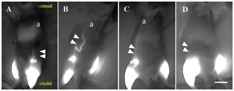
Fig 2. Representative images of lymphography after lymphatic ligation.
(A) Lymph flow obtained on the sham operation side on day 30. (B) Some detours were detected in the abdomen on day 30. These detours originated from the lower portion from the caudal ligation site (closed white arrowhead) of the collecting LV (a) and passed through the inguinal LN and connected to the upper portion from the cranial ligation (closed white arrowhead) site to collecting LV (a) passing through the inguinal LN. (C) An original route was detected after lymphatic ligation on day 30. (D) No lymph flow was detected on the operation side on day 18. a: the collecting LV that passed through the iliac LN and flowed into the axillary LN; scale bar of 1 cm; asterisk: inguinal LN; closed white arrowheads: ligation sites.

