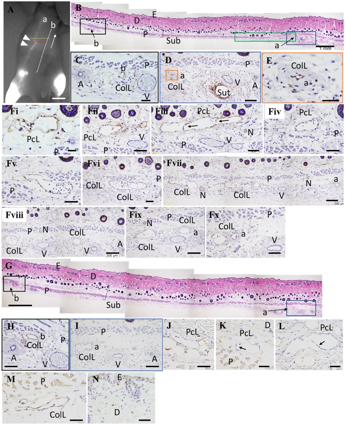
Fig 3. Representative histological images of detours at the proximal surgical site.
(A) Lymphography of the operation side on day 30. The red line is the starting point for the serial section. The yellow line is the end point for the serial section. The white arrow shows the direction of sectioning; scale bar of 1 cm. (B), (G) Hematoxylin and eosin (HE) staining. (C to F), (H), (I) Immunostaining with the anti-Podoplanin antibody. (B) The section at the red line in Fig 3A; scale bar of 1 mm. (C) A high magnification image in the black box in Fig 3B; scale bar of 50 μm. (D) A high magnification image in the blue box in Fig 3B. A transverse section of the 6–0 nylon suture (Sut) was observed; scale bar of 100 μm. (E) A high magnification image in the orange box in Fig 3D. A small lymphatic canal was observed and smooth muscle forming the wall of the ColL appeared to be thicker; scale bar of 50 μm. (Fi to Fx) A serial section of detours in the green box in Fig 3B. It is easy to identify the pre-collecting LV (PcL), stained in brown showing Podoplanin in the lymphatic endothelial cells, that gradually run from above the panniculus carnosus muscle (P) through the muscle to the ColL under the muscle. Many lymphocytes were present in the LVs (round cells stained with purple). Valves can be observed in the PcL (see arrows in Fig Fiii); scale bar of F(i), 20 μm; scale bar of Fii–Fx, 100 μm; scale bar of Fvii–Fix, scale bar of 200 μm. (G) The section at the yellow line in Fig 3A; scale bar of 1 mm. (H) A high magnification image in the black box in Fig 3G. There was a group of arteries (A), veins (V), and ColL (b) which were adjacent to the LV (a) in (A) shown using ICG; scale bar of 100 μm. (I) A high magnification image in the blue box in Fig 3G. Only ColL was observed near the collateral artery (A) and vein (V); scale bar of 200 μm. (J to N). Immunostaining to detect EdU. (J) A PcL with no EdU stain seen on day 15; scale bar of 40 μm. (K and L) PcLs with valves (arrows) with no EdU stain seen on day 30; scale bar of 40 μm. (M) A PcL with no EdU stain seen on day 30; scale bar of 40 μm. (N) Detection of EdU+ cells; epidermal cells; hair follicular cells, and; fibroblasts, stained brown in the epidermis and dermis; scale bar of 50 μm. a: LVs passed through the iliac LN and ran into the axillary LN; A: artery; arrows: valve of LV; asterisk: inguinal lymph node (LN); b: LVs that started at the lower abdomen and ran into the axillary LN; closed white arrowheads: ligation sites; ColL: collecting LV; D: dermis; E: epidermis; N: nerve; P: panniculus carnosus muscle; PcL: pre-collecting LV; Sub: subcutaneous tissue; Sut: suture; V: vein.

