Figure 4.
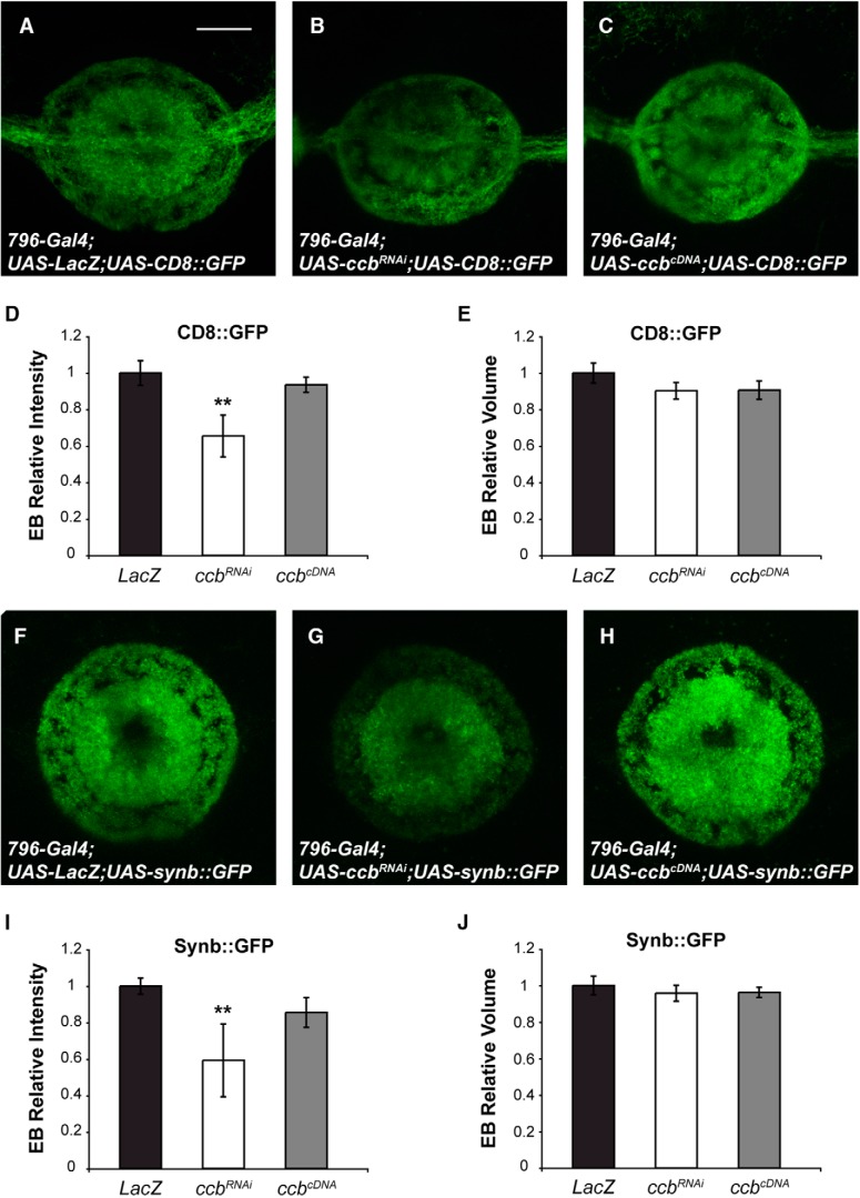
Axonal transport in adult R-neurons of the EB. Quantification of axonal transport of CD8::GFP (A–E) or Synaptobrevin::GFP (F–J) in R-neurons of the EB with downregulated or upregulated ccb. D, I, Axonal transport of CD8 and Synaptobrevin in R-neurons is shown as the average GFP intensity of CD8::GFP (D) and Synaptobrevin::GFP (I) in the EB and normalized to the GFP intensity in controls. Expression of the ccbRNAi in R-neurons significantly reduces axonal transport of CD8 (B; Kruskal–Wallis, p = 0.0072) and Synaptobrevin (G; Kruskal-Wallis, p = 0.0052) to the axon terminals in the EB. Expression of the ccbcDNA does not modify the transport ratio for CD8 (C; Kruskal–Wallis, p = 0.1381) or Synaptobrevin (H; Kruskal–Wallis, p = 0.5778). E, J, The total volume of the EB is measured for each individual expressing either CD8::GFP (E) or Synaptobrevin::GFP (J). The volume of the EB in CD8 expressing flies (E; Kruskal–Wallis, p > 0.9999 for flies coexpressing the ccbRNAi and p = 0.4518 for flies coexpressing the ccbcDNA) or in Synaptobrevin-expressing flies (J; Kruskal–Wallis, p > 0.9999 for flies coexpressing the ccbRNAi and p > 0.9999 for flies coexpressing the ccbcDNA) does not change. These results indicate that ccb levels do not alter the volume or morphology of the R-neurons and, thus, the effects on CD8 and Synaptobrevin signals are specific of their axonal traffic. Scale bar, 10 μm. Error bars indicate SEM. **p < 0.01; n = 6–14 per group.

